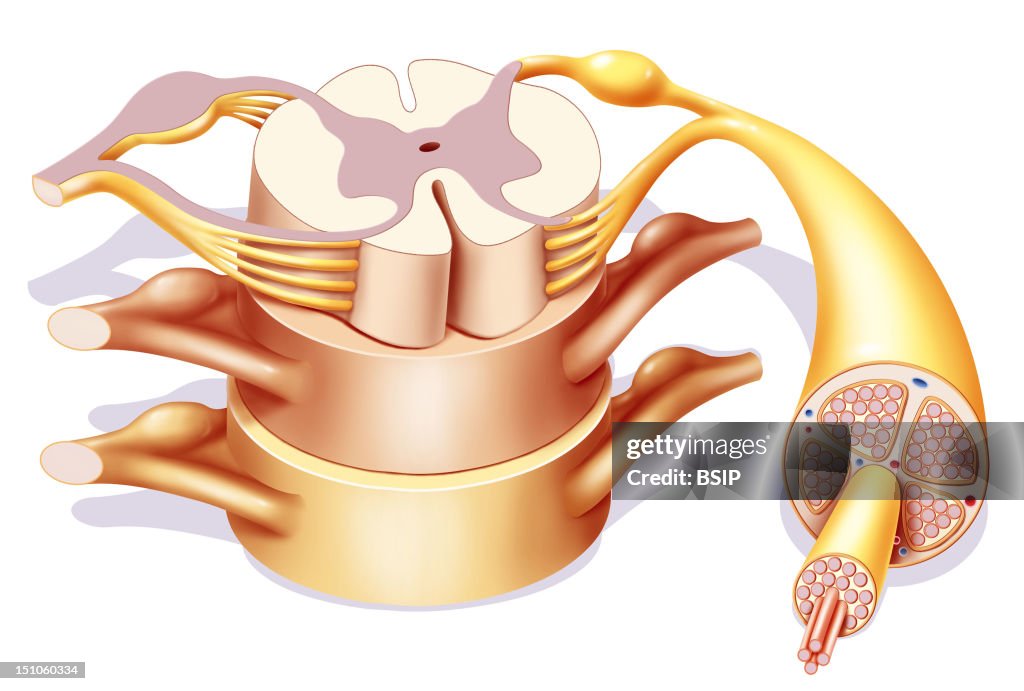
Spinal Cord, Drawing
The Spinal Cord. Representation In 3/4 Front View Of The Stucture Of The Spinal Cord, And Rachidian Nerves. From The Spinal Cord, Detach On Each Side Rachidian Nerves Constituted Of A Ganglion, A Posterior And An Anterior Root. A Cutaway View Of The Rachidian Nerve Highlights Its Structure With From The Outside Towards The Inside, The Epineurium In Which Are Located Its Blood Vessels, The Perineurium Surrounding The Isolated Nervous Fibers With An Effect In Zoom In. (Photo By BSIP/UIG Via Getty Images)

PURCHASE A LICENCE
How can I use this image?
$475.00
+GST AUD
Getty ImagesSpinal Cord, Drawing, News Photo Spinal Cord, Drawing Get premium, high resolution news photos at Getty ImagesProduct #:151060334
 Spinal Cord, Drawing Get premium, high resolution news photos at Getty ImagesProduct #:151060334
$650+GST$200+GST
Getty Images
In stockPlease note: images depicting historical events may contain themes, or have descriptions, that do not reflect current understanding. They are provided in a historical context. Learn more.
DETAILS
Restrictions:
Contact your local office for all commercial or promotional uses.
Credit:
Editorial #:
151060334
Collection:
Universal Images Group
Date created:
25 April, 2008
Upload date:
Licence type:
Release info:
Not released. More information
Source:
Universal Images Group Editorial
Object name:
941_04_1445608
Max file size:
3630 x 2449 px (30.73 x 20.73 cm) - 300 dpi - 1 MB