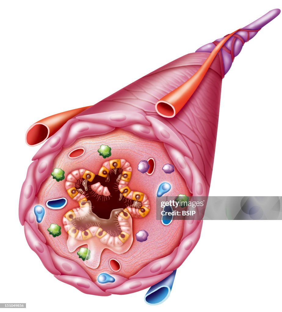
Asthma, Drawing
Asthmatic Bronchus. Illustration Of A Bronchus During An Asthma Attack. From The Outside Inwards: The Muscle Layer Is Hypertrophic, Immune Cells Mastocytes, Lymphocytes, Eosinophilic Leukocytes Are Present In The Inflamed Bronchial Mucosa And There Is Excessive Mucus Secretion With Desquamation Of The Epithelial Cells. The Combination Of These Factors Has Narrowed The Bronchial Lumen, Blocking The Passage Of Air. (Photo By BSIP/UIG Via Getty Images)

PURCHASE A LICENCE
How can I use this image?
$475.00
+GST AUD
Getty ImagesAsthma, Drawing, News Photo Asthma, Drawing Get premium, high resolution news photos at Getty ImagesProduct #:151049856
 Asthma, Drawing Get premium, high resolution news photos at Getty ImagesProduct #:151049856
$650+GST$200+GST
Getty Images
In stockPlease note: images depicting historical events may contain themes, or have descriptions, that do not reflect current understanding. They are provided in a historical context. Learn more.
DETAILS
Restrictions:
Contact your local office for all commercial or promotional uses.
Credit:
Editorial #:
151049856
Collection:
Universal Images Group
Date created:
03 October, 2005
Upload date:
Licence type:
Release info:
Not released. More information
Source:
Universal Images Group Editorial
Object name:
941_04_1172505
Max file size:
2979 x 3300 px (25.22 x 27.94 cm) - 300 dpi - 2 MB