Bladder cancer
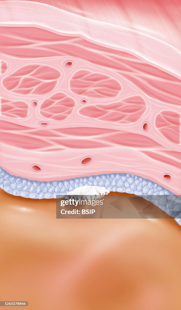
Stage 0a superficial bladder cancer. This illustration shows a zoom at the level of the bladder wall with all its structures. From bottom to top we have the bladder cavity, the epithelium (bluish gray cells), the connective tissue, the muscle consisting of three layers and finally the tissue that surrounds the bladder, here the parietal peritoneum. A superficial cancer tumor, or non-invasive papillary urothelial carcinoma is visible in this white drawing. (Photo by: BSIP/Education Images/Universal Images Group via Getty Images)

PURCHASE A LICENCE
How can I use this image?
$475.00
+GST AUD
Getty ImagesBladder cancer, News Photo Bladder cancer Get premium, high resolution news photos at Getty ImagesProduct #:1265278846
 Bladder cancer Get premium, high resolution news photos at Getty ImagesProduct #:1265278846
$650+GST$200+GST
Getty Images
In stockPlease note: images depicting historical events may contain themes, or have descriptions, that do not reflect current understanding. They are provided in a historical context. Learn more.
DETAILS
Restrictions:
Contact your local office for all commercial or promotional uses.
Credit:
Editorial #:
1265278846
Collection:
Universal Images Group
Date created:
08 May, 2020
Upload date:
Licence type:
Release info:
Not released. More information
Source:
Universal Images Group Editorial
Object name:
941_28_bsip_016055_006.jpg
Max file size:
3100 x 5320 px (26.25 x 45.04 cm) - 300 dpi - 3 MB